


Fibrotic myopathy in horses is a condition where fibrous scar tissue replaces normal muscle tissue, often in the hind limbs. This causes mechanical lameness and a classic gait abnormality known as “goose stepping.” Typically resulting from trauma or repeated intramuscular injections, the condition limits forward motion and is commonly seen in western performance horses.
What Is Fibrotic Myopathy?
Fibrotic myopathy is a performance-limiting condition characterised by the replacement of normal muscle fibers with fibrous scar tissue. This replacement typically occurs in the hamstring muscles of the hind limbs, particularly the semimembranosus and semitendinosus muscles. The resulting fibrotic tissue leads to mechanical restriction, hindering forward motion and creating a distinctive “goose stepping” gait abnormality. Though it’s not always painful, it significantly affects muscle function and can disrupt a horse’s normal gait.
How Muscle Tissue Is Affected
In fibrotic myopathy, injury to muscle tissue, often from trauma or repeated intramuscular injections, triggers a healing response where scar tissue forms in place of damaged muscle fibers. This scar tissue lacks the elasticity and contractile properties of normal muscle, causing fibrous tissue to bind parts of the muscle that should move freely.
This is especially problematic in the semitendinosus and semimembranosus muscles, where even mild cases can lead to lasting mechanical lameness and gait abnormalities. Deep palpation often reveals hardened areas along affected muscles.
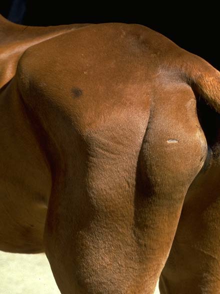
Image source: https://www.vetlexicon.com/equis/musculoskeletal/articles/muscle-myopathy-fibrotic/
Breed Susceptibility and Risk Profiles
Quarter Horses and other western performance horses are particularly susceptible to fibrotic myopathy. Their frequent use in sliding stops, sharp turns, and intense repetitive strain activities increases the likelihood of trauma to the hind limbs.
Chronic cases can also stem from poor injection techniques, especially repeated IM injections into hamstring muscles. Genetic predispositions or congenital abnormalities may also contribute, but repetitive motion and mechanical stress remain the most common culprits.
Recognizing Clinical Signs
Affected horses typically display a classic gait abnormality: the hind limb abruptly halts forward motion and slaps the hoof to the ground in a “goose stepping” motion.
This form of mechanical lameness is usually not painful in chronic cases but may show tenderness in acute conditions. During physical examination, clinical signs include shortened stride, abnormal limb movement, and palpable fibrotic bands in the hamstring region. Pain, muscle tension, or discomfort may accompany more acute cases.
Veterinary Diagnosis
Veterinarians diagnose fibrotic myopathy through a combination of physical examination and imaging. Deep palpation of the affected limb often reveals hardened, rope-like bands in the semimembranosus or semitendinosus muscle.
Diagnostic tools like ultrasound, thermography, or scintigraphy provide visual confirmation of fibrotic changes. In unclear cases, muscle biopsy may reveal the presence of scar tissue replacing normal muscle fibers. Diagnosis is typically based on clinical signs and confirmed through imaging or biopsy.
Affected Muscular Structures
The semitendinosus muscle and semimembranosus muscles are the most commonly affected in fibrotic myopathy. These hamstring muscles play a vital role in forward propulsion, and fibrous tissue in these areas severely limits function.
Occasionally, the biceps femoris or gracilis muscles may also be involved. Once fibrotic bands form, they create mechanical restriction, reducing muscle strength and altering limb dynamics. One hind limb is usually affected, but bilateral cases, though rare, have been reported.
Treatment Approaches
Treatment depends on severity and duration. Acute cases may respond to rest, NSAIDs, and cold therapy. For chronic or performance-limiting cases, physical therapy including therapeutic ultrasound, shockwave therapy, stretching exercises, and underwater treadmill therapy is often effective. Repeated IM injections in the affected muscles should be avoided. Surgical intervention, like standing myotomy, is considered when non-surgical methods fail to restore normal gait. Controlled exercise plays a pivotal role in rehabilitation.
Non-Surgical Options:
- Therapeutic ultrasound and shockwave therapy
- Controlled stretching exercises
- Underwater treadmill therapy
- Massage and manual therapy
- Progressive controlled exercise
Managing Acute Cases
In newly affected horses, the goal is to reduce inflammation and prevent fibrotic changes. This includes rest, NSAIDs, and cold compresses. Acute trauma should be addressed promptly, and muscle function monitored closely.
Therapeutic ultrasound and physical therapy may help mitigate early scar tissue formation. Stretching and light controlled exercise may begin once pain subsides. Identifying the initial injury and minimizing mechanical restriction can prevent progression to chronic cases.
Dealing with Chronic Conditions
Chronic cases often involve extensive fibrotic tissue, requiring longer-term strategies. Standing myotomy is a preferred surgical technique to release fibrous bands without general anesthesia. Horses with severe cases may show permanent gait abnormalities even after treatment.
However, physical therapy and hydrotherapy can improve muscle strength and limb mobility. Veterinary medicine continues to explore alternatives like laser fibrotomy and regenerative therapies to improve outcomes.
Rehabilitation and Recovery
Rehabilitation after surgery or therapy involves gradual reconditioning. Initial stall rest is followed by controlled walking, then trotting and cantering. Stretching exercises and therapeutic ultrasound support recovery of muscle function. Underwater treadmill therapy reduces strain while improving mobility. Daily monitoring of gait and muscle tone helps adjust the rehab program. Affected horses may require several months before returning to full work.
Recovery Strategy Includes:
- Passive range-of-motion exercises
- Gradual introduction of gaits
- Daily massage and ultrasound
- Hydrotherapy (if available)
Prognosis Based on Severity
The prognosis depends on how early the condition is diagnosed and treated. Mild cases may resume normal gait with conservative therapy. Severe cases with longstanding fibrotic bands often require surgery, with about 50% of horses returning to prior performance levels.
Prognosis improves with dedicated rehabilitation and proactive management. Chronic or untreated fibrotic myopathy may become performance-limiting, especially in competitive settings.
Prevention and Best Practices
Preventing fibrotic myopathy involves minimizing trauma to hind limbs and maintaining muscular health. Limit intramuscular injections into hamstring muscles, especially in performance horses. Always warm up and cool down properly, and avoid abrupt maneuvers on poor footing. Controlled exercise and regular stretching reduce repetitive strain. For horses with previous injury, preventive therapies and vet monitoring can reduce recurrence.
Prevention Tips:
- Use alternative injection sites when possible
- Ensure proper warm-up and cool-down
- Avoid repetitive high-strain maneuvers
- Include stretching in daily routines
- Provide adequate turnout and conditioning
Data Overview
[Fibrotic Myopathy in Horses: Key Comparisons and Facts]
| Aspect | Summary |
|---|---|
| Definition & Mechanism | Scar tissue replaces muscle, especially in hamstrings, causing “goose stepping” gait |
| Affected Muscles | Mainly semitendinosus and semimembranosus |
| Primary Causes | Trauma, repetitive strain, IM injections |
| Clinical Signs | Goose stepping, mechanical lameness, palpable fibrous bands |
| Diagnosis Tools | Physical exam, deep palpation, ultrasound, biopsy |
| Non‑surgical Treatment | Ultrasound, underwater treadmill, stretching |
| Surgical Options | Standing myotomy, tenotomy, tissue excision |
| Rehabilitation | Controlled exercise, ROM work, hydrotherapy |
| Prognosis | Mild cases recover well, 50% return after surgery |
| Prevention | Avoid injections, use warm-ups, maintain muscle fitness |
Source: Veterinary medicine sources including AAEP, vetmed publications, and peer-reviewed journals.
Typical Questions
Below are typical questions we get asked about this topic.
What causes fibrotic myopathy in horses?
Fibrotic myopathy typically develops from muscle damage—often due to trauma, repetitive strain, or repeated intramuscular injections in the hind limbs. It’s especially common in western performance horses like Quarter Horses performing sharp turns or sliding stops.
Which muscles are most affected?
The semimembranosus and semitendinosus muscles/tendons are most commonly involved. Scar tissue forms in these hamstring muscles, creating mechanical restriction during limb extension.
What are the clinical signs to look for?
Signs include:
– A “goose stepping” gait, hind hoof slaps down instead of a normal stride.
– Mechanical lameness, no true pain once it becomes chronic.
– On examination, hardened bands of fibrous tissue may be felt via deep palpation in acute cases.
How is the condition diagnosed?
Diagnosis is based on:
– Clinical signs and physical examination.
– Deep palpation to identify fibrous bands.
– Imaging (ultrasound, thermography, scintigraphy).
– Biopsy in ambiguous cases, showing fibrotic replacement of muscle tissue.
Can fibrotic myopathy be treated without surgery?
Yes. Non‑surgical options include:
– Controlled exercise, progressive walk, trot, canter.
– Stretching exercises, therapeutic ultrasound, hydrotherapy, massage, shockwave, and underwater treadmill therapy.
– NSAIDs and cold/hot therapy in acute cases to address pain and inflammation.
When is surgery recommended?
Surgery is considered for severe cases or when conservative treatment fails. The common procedure is a standing myotomy, where the fibrotic band is cut under sedation. Tenotomy or laser fibrotomy may also be used to remove or break up scar tissue.
What does recovery and rehabilitation involve?
Post-treatment recovery includes:
– Stall rest with passive range-of-motion exercises.
– Gradually increasing to controlled exercise (walk → trot → canter).
– Continued therapies like hydrotherapy or ultrasound, and careful monitoring of muscle strength and gait improvement.
What is the prognosis long-term?
For mild or chronic cases, horses may continue performing normally with conservative management.
After surgery, approximately 50% of athletic horses regain intended performance, though recurrence is possible, especially if prevention practices aren’t followed.
How can I prevent fibrotic myopathy in my horse?
Prevention strategies include:
– Gradual warm-up and cool-down routines.
– Avoiding muscle injections in the hind limbs when possible.
– Ensuring good footing and avoiding abrupt athletic maneuvers like sliding stops or tight turns.
– Maintaining consistent conditioning, turnout, and strength work to build muscle resilience.


